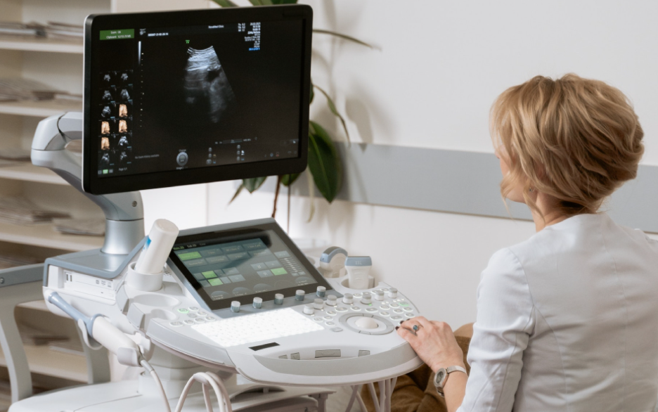
Noninvasive Cardiovascular Sonography, also known as echocardiography, uses sound waves to create images of the heart and blood vessels. The procedure is performed using a device called a transducer, which emits high-frequency sound waves that bounce off the structures of the heart and blood vessels. The echoes are then captured and translated into images by a computer, which can be viewed on a monitor.
During the procedure, the patient will lie on an exam table and gel will be applied to the chest area to improve the transmission of sound waves. The diagnostic medical sonographer will then move the transducer over the chest area, capturing images of the heart and blood vessels from different angles. The procedure is usually done in a hospital, clinic or doctor’s office, and it is non-invasive, painless and does not require any special preparation.
The images obtained from Noninvasive Cardiovascular Sonography can provide detailed information about the size, shape, and function of the heart and blood vessels, including information about the heart’s chambers, valves, blood flow, and blood flow through the blood vessels. This information can be used to diagnose and monitor a variety of cardiovascular conditions, such as heart disease, hypertension, and valvular heart disease.
Principles of ultrasound technology in Noninvasive Cardiovascular Sonography
The principles of ultrasound technology in Noninvasive Cardiovascular Sonography involve the use of high-frequency sound waves to create images of the heart and blood vessels.
Ultrasound waves, or sound waves with a frequency higher than the upper limit of human hearing, are emitted by a device called a transducer. The transducer sends out sound waves that travel through the skin and tissue and bounce off the structures of the heart and blood vessels. The echoes are then captured by the transducer and sent to a computer, which converts them into images that can be viewed on a monitor.
The speed of sound is different in different types of tissue, and this difference allows the noninvasive cardiovascular sonographer to create images of the different structures within the heart and blood vessels. For example, blood, which is more dense than muscle or fat, will reflect sound waves differently, creating a bright area on the image. The computer can also use the Doppler effect, which is the change in frequency and wavelength of a wave in relation to the movement of the source or the observer, to show the direction and velocity of blood flow.
Noninvasive Cardiovascular Sonography is a very safe procedure, it does not use ionizing radiation and is not associated with any known long-term risks or side effects.
The technique is very versatile, it can be used to evaluate the structure and function of the heart, including the size and shape of the heart’s chambers, the function of the heart’s valves, the amount of blood flowing through the heart and blood vessels, and the presence of any abnormal masses or fluid collections in the heart or blood vessels.
Types of Noninvasive Cardiovascular Sonography
There are several different types of Noninvasive Cardiovascular Sonography, each with its own set of benefits and applications.
- Transthoracic echocardiography (TTE): This is the most common type of echocardiography and it is performed by placing a transducer on the patient’s chest. TTE is used to evaluate the overall function of the heart, including the size and shape of the heart’s chambers, the function of the heart’s valves, and the amount of blood flowing through the heart.
- Transesophageal echocardiography (TEE): This type of echocardiography is performed by placing a transducer in the patient’s esophagus, allowing for more detailed images of the heart, especially the back of the heart and the blood vessels entering and leaving the heart. TEE is often used to evaluate the heart for blood clots, tumors, or other abnormalities.
- Stress echocardiography: This type of echocardiography is performed while the patient is exercising or taking medication to increase the heart rate, which allows the cardiologist to assess how the heart functions under stress. Stress echocardiography is used to evaluate patients with suspected or known coronary artery disease.
- Doppler echocardiography: This type of echocardiography uses the Doppler effect to measure the speed and direction of blood flow through the heart and blood vessels. Doppler echocardiography is used to evaluate patients with suspected or known blood flow problems, such as aortic stenosis.
All these types of Noninvasive Cardiovascular Sonography can be performed in a hospital, clinic or doctor’s office and usually only take about 30 minutes to complete.
Conclusion
In conclusion, Noninvasive Cardiovascular Sonography is a safe and effective technique for evaluating the structure and function of the heart and blood vessels. It uses high-frequency sound waves to create images of the heart and blood vessels, and it does not use ionizing radiation or cause any known long-term risks or side effects. There are several different types of Noninvasive Cardiovascular Sonography, including transthoracic echocardiography, transesophageal echocardiography, stress echocardiography, and Doppler echocardiography. Preparing for the procedure is simple and straightforward, and it usually only takes about 30 minutes to complete. It is an essential tool for the diagnosis, treatment, and management of cardiovascular diseases.